Explore pterygium removal surgery options with GetWellGo. Learn about causes, symptoms, and treatment for international patients seeking top-quality eye care.
Pterygium surgery is the process where a pterygium lies on the cornea is removed. This mainly arises from the sun exposure, constant scrubbing or exposure to wind, and dryness of the eye. The surgery is usually done if:
It obstructs vision due to its growth on the facial skin specifically the cornea.
Causes discomfort or recurrent inflammation
Is cosmetically concerning
Pterygium Surgery
Pterygium surgery is a minor operation to remove a ‘wing-like’ growth of conjunctiva tissue could extend on the cornea. Surgery is usually recommended when the pterygium:
Affects or threatens vision
Causes chronic irritation or inflammation
Is cosmetically disfiguring
Recurs or grows rapidly
Types of Pterygium Surgery
Bare Sclera Excision
Simple excision of the pterygium with the white part of the eye sclera being exposed
High recurrence rate (30–80%)
Conjunctival Autograft (Preferred Method)
Following pterygium excision, conjunctival autograft, most probably from the upper part of the eye) is used to cover the area.
Low recurrence (2–10%)
Usually choose between applying fibrin glue and suturing to affix the graft.
Amniotic Membrane Transplant
This approach is used when the continent of autografts is not possible, for example if the area has had previous surgeries.
Little recurrence rates that make it appropriate in recurrent cases or extensive pterygia.
Adjunctive Therapy with Mitomycin C or 5-Fluorouracil (5-FU)
Patients given preoperatively, intraoperatively or postoperatively in order to decrease the chances of reformation of fibrosis
Should be used cautiously due to possible side effects such as deterioration of scleral thickness
Pterygium Treatment
The decision of managing pterygium depends with the size of the lesion and the clinical manifestation as well as the behavior of the lesion. Simple pterygium might not need surgery, yet an extensive or invasive lesion most probably will require surgical treatment.
Non-Surgical Treatment (For Mild Cases)
Recommended when pterygium is minimal, stable and not exerting influence on the patient’s vision.
Lubricating eye drops and artificial tears
They include reduction in dryness, surface irritation and foreign body sensation.
As for the long-term therapy, it is recommended to use preservative-free artificial tears.
Anti-Inflammatory Drops
Some of the enabling aggravating symptoms include: Mild Steroid or NSAID eye drops help to reduce redness and swelling.
It is used in the short-term treatment in order to minimize side effects such as elevation of intraocular pressure.
UV and Dust Protection
Wear wraparound sunglasses to block UV rays
Avoid windy or dusty environments
Pterygium Eye Surgery
Pterygium eye surgery is a surgical operation that aims at removing pterygium which is a wing-shaped growth of conjunctival tissue that can grow over the cornea due to exposure to UV light, dusty and dry conditions.
Why Pterygium Surgery Is Done?
Surgery is recommended when:
It is occupying the more central area together with growing further towards the central part of the cornea and with impact on vision.
This leads to inflammation, skin rash, or soreness or the skin, eyes, or mucous membrane.
There's significant cosmetic concern
It is a recurrent or a giant pterygium
Factors Affecting Pterygium Removal Cost
The cost of pterygium operation depends on a number of factors, which can even differ for different countries, clinics, surgeons, and other peculiarities.
Risk, Recurrence and Counselling: Before engaging in any operation, the patient should consent to it, and the surgeon must explain the possible risks, the recurrence of certain conditions and any post-operation care to the patient.
Anaesthesia: Local anaesthetic eye drops or injection; mild sedation in some cases
Surgical Procedure Steps
Step 1: Pterygium Excision
The pterygium tissue is then lifted and taken out from the corneal and scleral surfaces by the hand of the surgeon carefully.
Step is taken to avoid trauma to the surrounding healthy tissue of the cornea during the cleaning processes.
Step 2: Graft Preparation
A conjunctival autograft is obtained from the superior part of the eye which may be located behind the upper eyelid.
In the instances when autograft cannot be incorporated (for example when the surgery is the second one), an amniotic membrane may be used.
Step 3: Graft Placement
The pterygium is then trimmed and excised, and the graft is sown over the area.
It is fixed using either:
Fibrin glue which is more comfortable and has a quicker recovery time than other forms of hemostasis.
Sutures (absorbable or non-absorbable but may lead to mild irritation)
Step 4: Adjunctive Therapy (if needed)
The use of Mitomycin C (MMC) on patients in order to reduce the recurrence risk, most especially on cases that are tagged as aggressive or recurrent.
Duration
It is normally done within a period of 30 to 60 minutes depending on the formulation used and the level of diffusion.
Postoperative Protocol
Therefore temporary use of eye patch or shield can be worn for some number of hours or overnight.
Antibiotic with steroid eye drops as prescribed for 2-4 weeks
Do not rub the eye, or perform activities that require heavy lifting or lifting weights and stay away from direct sun exposure especially during the summer times.
The follow-ups are usually made at Day 1, Week 1, and on monthly basis
Pterygium Removal Before and After
Before Surgery
Appearance:
Sore associated with a mass tumor of bright pink fleshy structure originating from the white part of the eye, conjunctiva and protruding onto the space occupied by the cornea.
May appear triangular or wing-shaped
Eye may appear to be red in color, looking inflamed, or may seem to be dry.
Symptoms:
Foreign body sensation
Chronic redness or inflammation
Distortion of vision while seeing (if growth extends up to corneal center)
Cosmetic concern or self-consciousness
Pre-op Care:
Eye exam with slit-lamp
Presumably, patients should avoid using contact lenses prior to the surgery.
Application of drops on the eyes and putting on sunglasses to reduce the irritation.
After Surgery
Immediately After:
Redness, swelling, and mild irritation
Lesions in the graft area may appear erythematosus and slightly elevated
Eye patch or shield for the first day
Vision may be temporarily blurred
Appearance (1–4 Weeks):
It fuses with the natural graft tissue after several hours of operation
Redness gradually subsides
The eyes have become clearer and usual sclera transparency is seen.
Cosmetic improvement is often significant
Long-Term (2–3 Months):
Eye returns to normal appearance
Vision typically stabilizes
No visible growth if the tumor is interpreted to mean that it has not regrown, after treatment.
This depends with the kind of scarring that would be incurred in the event that the healing process is effective.
Pterygium Surgery Risks
Pterygium surgery is relatively innocuous however they are associated with some risks and probable complications as is the case with most surgical intervention. It is useful to be aware of these to make a good decision and avoid mistaking something related to the post-operation care.
Pterygium surgery complications:
Recurrence of Pterygium
Redness and Irritation
Graft-Related Issues
Infection
Corneal Scarring or Thinning
Double Vision (Diplopia) or Eye Movement Problems
Allergic Reactions
Pterygium Removal Success Rate
Bare Sclera Excision ~20–70% (high recurrence rate)
With Mitomycin C (MMC) Often improves success, but risks must be weighed
Pterygium Surgery Outcomes
Pterygium surgery in general yields satisfactory results if the latest method of conjunctival autograft is applied. The following is a brief on the likely outcomes after surgery.
Positive Outcomes:
Cosmetic Improvement
Visual Improvement
Low Recurrence (with advanced techniques)
Quick Recovery
Why Choose GetWellGo for Pterygium Surgery?
GetWellGo is regarded as a leading supplier of healthcare services. We help our foreign clients choose the best treatment locations that suit their needs both financially and medically.
We offer:
Complete transparency
Fair costs.
24 hour availability.
Medical E-visas
Online consultation from recognized Indian experts.
Assistance in selecting India's top hospitals for pterygium surgery.
Expert ophthalmologist with a strong track record of success
Assistance during and after the course of treatment.
Language Support
Travel and Accommodation Services
Case manager assigned to every patient to provide seamless support in and out of the hospital like appointment booking
Celebrate a historic milestone of 2500 successful Bone Marrow Transplant at Fortis Gurgaon procedures, backed by our complete international patient visa care.
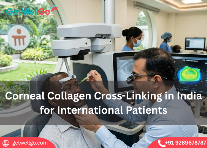
Experience advanced Corneal Collagen Cross-Linking in India to stabilize keratoconus and protect your sight, with full medical visa help from our travel team.
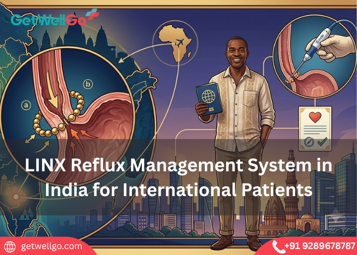
Experience the LINX Reflux Management System in India to stop severe heartburn using a tiny magnetic ring, fully organized by our fast-track medical visa team.
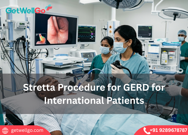
Experience advanced Stretta Procedure for GERD in India to eliminate chronic acid reflux without surgery, completely managed by our fast-track medical visa team
Medical tourism in India for Uzbekistan Patients made easy. GetWellGo handles your top-tier surgery, Uzbek translator, and a safe, warm home for your family.
Palliative care services in India focus on your comfort and peace of mind. GetWellGo handles the expert pain relief, visa, and a serene, private home to heal.
Transjugular Intrahepatic Portosystemic Shunt in India stops dangerous liver bleeding. GetWellGo handles your top specialist, visa, and a safe, cozy stay.
Keratoconus Treatment in India stops your vision from getting worse. GetWellGo handles your advanced eye care, visas, and a safe trip to save your sight.
Implantable Collamer Lens in India lets you ditch thick glasses forever. GetWellGo handles your expert eye doctor, flights, and a clear-vision holiday.
Cochlear Implants for Children in India opens a world of sound. We at GetWellGo plan your affordable ear surgery, airport pickup, and safe family trip.