
Historic Milestone: FMRI Celebrates 2500 Successful Bone Marrow Transplants
Celebrate a historic milestone of 2500 successful Bone Marrow Transplant at Fortis Gurgaon procedures, backed by our complete international patient visa care.
Read MoreGetWellGo provides a comprehensive guide on VP shunts, detailing types, surgical procedures, and recovery insights for patients seeking information on hydrocephalus treatment

The surgery may take an average of 1 to 2 hours and requires general anaesthesia.
Incisions:
Placement of Catheter:
Valve Insertion:
Connection and Securing:
Closure:
Wounds are then sutured or stapled, and cleaned, dressed with sterile materials.
Ventriculoperitoneal Shunt is an operation for managing conditions that cause increased levels of fluid in the brain called cerebrospinal fluid. This condition, known as hydrocephalus, causes higher pressure inside the skull so that patients may experience headaches, nausea, vision difficulties, or problematic brain function in the worst-case scenario.
The Ventriculoperitoneal Shunt (VP Shunt) procedure is long and characterized by minimal risk, although, like any surgery, it has some risks. These risks are associated with the procedure of implantation of the shunt as well as the further work of the shunt system. Knowledge of these risk factors plays role in identifying possible complication early enough to prevent.
The length of time it takes for someone who has undergone a ventriculoperitoneal (VP) shunt surgery to recover is different from one person to another, based on their general health, the reasons why the surgery was done, and whether the patient developed some complications. Here’s a general timeline:
Immediate Recovery (Hospital Stay)
Short-Term Recovery (First Few Weeks)
Long-Term Recovery (Full Healing)
VP shunt infection is a severe condition that should be handled by a doctor as soon as possible. There are some differences of symptoms depending of if the infection is localized in the shunt system, spread into the brain (meningitis) or if it affected the peritoneal cavity (peritonitis).
VP Shunt Placement
VP Shunt Malfunction Signs
Despite the effectiveness of ventriculoperitoneal shunting, there are certain risks factor that may arise with the surgery. They can present early or late after shunt surgery, may be related to an infection and may need shunt adjustment.
A VP shunt failure is a condition where there is failure of the shunt to drain peritoneal fluid to the ventricle as a result of blockage, infection, mechanical problems or inadequate functioning. This leads to the recurrence of hydrocephalus symptoms and a need for a quick doctor’s intervention.
VP shunt removal is a surgical procedure in which a part or whole of a ventriculoperitoneal shunt is removed. This is usually done if the shunt is not required any longer, is infected or is not working properly.
Reasons for VP Shunt Removal
Ventriculoperitoneal (VP) shunt surgery cost depends on many factors such as the hospital, the surgeon fees as well as post-operative management. Thus, the cost estimate consolidation takes into account the following factors:
Hospital Stay:
Imaging:
Physical Activity:
Follow-up Care
Monitoring Shunt Function:
An adjustment of the VP shunt is necessary when its pressure needs to be altered to effectively manage the drainage of cerebrospinal fluid (CSF). This is particularly for programmable shunts that include non-invasive pressure changes through a magnetic instrument.
GetWellGo is regarded as a leading supplier of healthcare services. We help our foreign clients choose the best treatment locations that suit their needs both financially and medically.
We offer:
Explore expert medical insights, treatment guides, and the latest healthcare updates for international patients - visit our blog .
Celebrate a historic milestone of 2500 successful Bone Marrow Transplant at Fortis Gurgaon procedures, backed by our complete international patient visa care.
Read More
Experience advanced Corneal Collagen Cross-Linking in India to stabilize keratoconus and protect your sight, with full medical visa help from our travel team.
Read More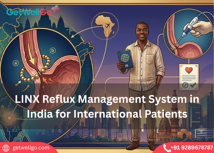
Experience the LINX Reflux Management System in India to stop severe heartburn using a tiny magnetic ring, fully organized by our fast-track medical visa team.
Read More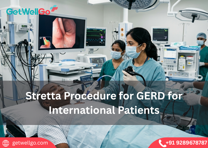
Experience advanced Stretta Procedure for GERD in India to eliminate chronic acid reflux without surgery, completely managed by our fast-track medical visa team
Read More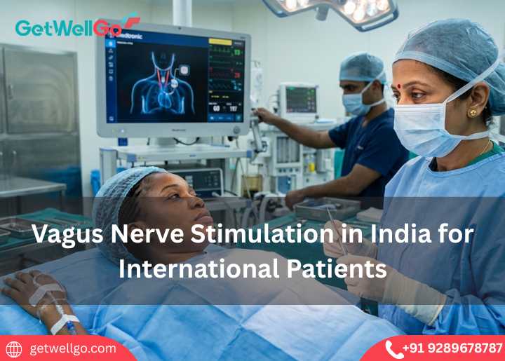
Get advanced vagus nerve stimulation treatment in India with expert care, modern hospitals, and affordable options for international patients.
Read More
Medical tourism in India for Uzbekistan Patients made easy. GetWellGo handles your top-tier surgery, Uzbek translator, and a safe, warm home for your family.
Read More
Palliative care services in India focus on your comfort and peace of mind. GetWellGo handles the expert pain relief, visa, and a serene, private home to heal.
Read More
Get reliable visa assistance for medical travel to India. Support with documentation, approvals, and a smooth entry process for global patients.
Read More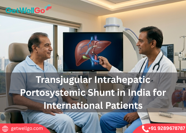
Transjugular Intrahepatic Portosystemic Shunt in India stops dangerous liver bleeding. GetWellGo handles your top specialist, visa, and a safe, cozy stay.
Read More
Keratoconus Treatment in India stops your vision from getting worse. GetWellGo handles your advanced eye care, visas, and a safe trip to save your sight.
Read More
Implantable Collamer Lens in India lets you ditch thick glasses forever. GetWellGo handles your expert eye doctor, flights, and a clear-vision holiday.
Read More
Cochlear Implants for Children in India opens a world of sound. We at GetWellGo plan your affordable ear surgery, airport pickup, and safe family trip.
Read More