Hydronephrosis: Causes, Symptoms, Treatment and Diagnosis
Learn about hydronephrosis causes, symptoms, diagnosis & treatment. Expert care at GetWellGo for international patients. Start your recovery journey today.
Hydronephrosis is a condition where the one or both kidneys swell up because of the accumulation of urine. It occurs when there is blockage or obstruction in the urinary tract that does not allow urine to drain normally from the kidney to the bladder.
Hydronephrosis causes
Hydronephrosis is a condition where the urine is unable to drain adequately from the kidney into the bladder, and the kidney becomes swollen. The etiology can be divided into location (obstructive vs non-obstructive), duration (acute vs chronic), and age group (infants vs adults).
Major Causes of Hydronephrosis
Obstruction in the Urinary Tract
These are the most frequent causes:
Kidney stones
Prevent urine flow from the kidney to the bladder.
Ureteral stricture
Ureter narrowing due to trauma, surgery, or inflammation.
Tumors (benign or malignant)
In the kidney, ureter, bladder, or nearby areas compressing on the urinary tract.
Benign prostatic hyperplasia (BPH) – in males
Compresses on the urethra and blocks the flow of urine.
Bladder outlet obstruction
Caused by tumors, stones, or functional issues.
Vesicoureteral Reflux (VUR)
Urine moves in a backward direction from the bladder to the ureters and kidneys.
Common in infants and children, usually diagnosed with a VCUG test.
Pregnancy
The expanding uterus may press the ureters, particularly the right one.
Typically temporary and improves after childbirth.
Congenital (At Birth)
Ureteropelvic junction (UPJ) obstruction
Obstruction where the ureter joins the kidney.
Posterior urethral valves (in male babies)
Tissue folds in the urethra that impede urine flow.
Neurogenic Bladder
Nerve dysfunction (e.g., in spinal cord injury, multiple sclerosis, or diabetes) may cause incomplete emptying of the bladder, resulting in backflow.
Ureterocele
A balloon-like distension of the lower ureter within the bladder, usually congenital, which obstructs flow of urine.
Post-Surgical or Post-Radiation Changes
Fibrosis or inflammation from operations or pelvic radiation can obstruct the urinary tract.
Hydronephrosis symptoms
Hydronephrosis symptoms vary from mild to severe and may either be dependent on whether the condition is chronic or acute (gradual or sudden), as well as whether one or both kidneys are involved.
Adult Symptoms
Pain in flank or back
Typically on the same side as the involvement
Sharp or dull
Painful abdomen or pressure
Dysuria (painful urination)
Frequent urination
Trouble urinating or failure to empty
Hematuria (blood in urine)
Nausea and vomiting
Fever and chills (if infected)
Cloudy or odor urine
Infant and Child Symptoms
Unexplained fever
Poor feeding
Fussiness or crying during urination
Vomiting
Abdominal distention
Failure to gain weight
Can be seen prenatally on ultrasound
Hydronephrosis treatment
Hydronephrosis treatment is aimed at eliminating the root cause of the urine obstruction and maintaining kidney function. The technique used is based on:
Severity (mild, moderate, or severe)
Its acute or chronic nature
Underlying cause (e.g., stone, tumor, pregnancy)
Patient's age and general health
Treat the Root Cause
Kidney Stones
Small stones: Can pass spontaneously; increased fluids + pain medications.
Large or obstructed stones:
Ureteroscopy
Lithotripsy (ESWL) – shatters the stone with sound waves
Percutaneous nephrolithotomy (for extremely large stones)
Infection (e.g., pyelonephritis or UTI)
Antibiotics to manage infection
Drainage may be required if infection is associated with blockage
Tumors or Prostate Enlargement
Surgery, chemotherapy, or radiation (depending on type)
Prostate treatment (e.g., medications or TURP surgery)
Pregnancy-related Hydronephrosis
Generally resolves after delivery
Pain management and observation unless complications develop
Congenital Blockages (in children)
Observation if mild and kidney function is normal
Surgery (such as pyeloplasty) if obstruction is severe or kidney function deteriorates
Relieve the Obstruction (Drainage Procedures)
Employed when urine cannot drain spontaneously:
Ureteral stent
Small tube inserted into the ureter to maintain patency
Nephrostomy tube
Tube inserted directly into the kidney to drain urine out of the body
Ongoing Monitoring (If Mild)
Ultrasound or CT scans to monitor kidney size and function
Blood tests (such as creatinine, BUN)
Watchful waiting can be applied during pregnancy or in mild conditions
Hydronephrosis in adults
Adult hydronephrosis is the inflation of a single or both kidneys with urine due to urine accumulation when there is a blockage or inadequate flow within the urinary tract. It's either acute (sudden) or chronic (insidious), and may occur in one (unilateral) or both kidneys (bilateral).
Hydronephrosis in pregnancy
Hydronephrosis in pregnancy is the enlargement of the kidney because of the accumulation of urine when its passage is being partially obstructed. It is a frequent condition during pregnancy and is normally physiological (normal) in nature but not pathological.
How Common Is It?
Occurs in up to 90% of pregnant women, particularly in the second and third trimesters.
More frequent on the right side owing to the position of the uterus and anatomical variations in the ureters.
Causes of Hydronephrosis during Pregnancy
Physiological
Hormonal changes: Progesterone causes relaxation of muscles of the ureters, reducing the flow of urine.
Uterine pressure: The expanding uterus presses against the ureters, particularly on the right side.
Pathological
Kidney stones
Urinary tract infections (UTIs)
Ureteral obstruction secondary to other causes
Hydronephrosis diagnosis
Hydronephrosis is diagnosed by imaging tests that show dilation (enlargement) of the renal pelvis and calyces, usually in conjunction with clinical symptoms or signs. Diagnosis seeks to establish the diagnosis and determine the cause of urinary tract obstruction.
Step-by-Step Diagnostic Approach
Medical History and Physical Exam
Symptoms: Flank pain, urinary symptoms, fever, etc.
Risk factors: Kidney stones, recent surgeries, pregnancy, history of UTIs
Physical exam may include:
Flank tenderness
Palpable kidney (rare)
Imaging Studies (Mainstay of Diagnosis)
Ultrasound (US)
First-line test
Identifies kidney swelling and severity
No radiation—safe in pregnancy and children
CT Scan (Non-contrast if stones suspected)
More detailed than ultrasound
Best for identifying:
Kidney stones
Tumors
Ureteral obstruction
Used if the ultrasound is unclear or complications are suspected
MRI / MR Urography
Alternative to CT if radiation has to be avoided
Especially helpful in pregnancy
Intravenous Pyelogram (IVP)
Earlier method; seldom used nowadays
X-ray imaging following an injection of dye to outline urinary tract
Urine Tests
Urinalysis:
Seek blood, white blood cells, bacteria, or protein
Urine culture:
When infection is suspected
24-hour collection of urine (in selected situations):
To assess stone risk
Blood Tests
Creatinine and Blood Urea Nitrogen (BUN):
Gauge kidney function
Electrolytes:
Check for imbalance due to decreased kidney function
Nuclear Renal Scan (Renography)
Reviews function of each kidney
Used to estimate severity of obstruction
Usually employed in chronic or complicated cases
Hydronephrosis surgery
Surgery may be necessary for hydronephrosis if there is a chronic or severe obstruction of the urinary tract, particularly if kidney function is in jeopardy or symptomatology is severe. The surgery varies with the cause, location of obstruction, and whether acute or chronic.
Surgical Choices:
Ureteral stent placement
Indication: Short-term bypassing of obstruction and enabling urine drainage from kidney to bladder.
Method: Thin plastic tube inserted within the ureter.
Used in:
Stones
Tumors
Inflammatory obstruction
Typically inserted endoscopically (cystoscopy) with anaesthesia.
Percutaneous Nephrostomy
Purpose: Relieves urine by draining directly from kidney to outer bag.
Method: Catheter inserted through the skin into renal pelvis.
Used when:
Ureter fully obstructed
Acute drainage is required (e.g., infection, sepsis)
Often temporary measure prior to definitive surgery.
Long-term solution, particularly in congenital or chronic conditions
Ureteral Reimplantation
Purpose: Repair obstruction or reflux at bladder end of ureter.
Method: Reattach ureter to a new location on the bladder.
Used in children with vesicoureteral reflux or narrowing of ureter
Stone Removal Procedures
If hydronephrosis is caused by kidney or ureteral stones, these can be removed surgically:
Ureteroscopy with laser lithotripsy
Percutaneous nephrolithotomy (PCNL) for large stones
Shock wave lithotripsy (SWL) (non-invasive)
Tumor Resection or Bypass
For cancer or extrinsic compression (e.g., due to a tumor in the pelvis), options are:
Tumor removal
Ureteral bypass
Palliative stenting or nephrostomy if surgery is not feasible
Hydronephrosis complications
Kidney Damage or Kidney Failure
Recurrent Urinary Tract Infections (UTIs)
Urosepsis
Kidney Stones
Hypertension (High Blood Pressure)
Electrolyte Imbalances
Urinary Incontinence or Retention
Pain and Discomfort
Ureteral obstruction and hydronephrosis
Ureteral obstruction is one of the main reasons for hydronephrosis. It happens when there is an obstruction in the flow of urine from the bladder to the kidney, resulting in urine accumulation and kidney swelling (hydronephrosis).
What Is Ureteral Obstruction?
Blockage or constriction of the ureter (the tube that leads each kidney to the bladder), which cuts off normal drainage of urine.
How It Causes Hydronephrosis
When the ureter becomes blocked:
The urine is not able to drain normally from the kidney.
Urine collects in the renal pelvis (the drainage component of the kidney).
It leads to increased pressure, enlargement, and swelling — the signature of hydronephrosis.
Why Choose GetWellGo for Hydronephrosis Treatment?
GetWellGo is regarded as a leading supplier of healthcare services. We help our foreign clients choose the best treatment locations that suit their needs both financially and medically.
We offer:
Complete transparency
Fair costs.
24 hour availability.
Medical E-visas
Online consultation from recognized Indian experts.
Assistance in selecting India's top hospitals for hydronephrosis treatment.
Expert urologist/nephrologist with a strong track record of success
Assistance during and after the course of treatment.
Ensure a safe recovery with a Pediatric Pyeloplasty in India organized smoothly by our team, from rapid child specialist booking to comfortable home stays.
Regain complete control with an Artificial Urinary Sphincter in India organized smoothly by our team, from rapid specialist booking to a cozy recovery stay.
Regain normal urinary flow with a Thulium Laser Prostatectomy in India organized smoothly by our team, from rapid specialist booking to a safe recovery stay.
Experience advanced Flexible Ureteroscopy in India to dust kidney stones quickly without incisions, fully supported by our medical visa and travel team.
Protect your quality of life using the advanced UroLift System in India with our dedicated care team handling your hospital bookings, visas, and warm stay.
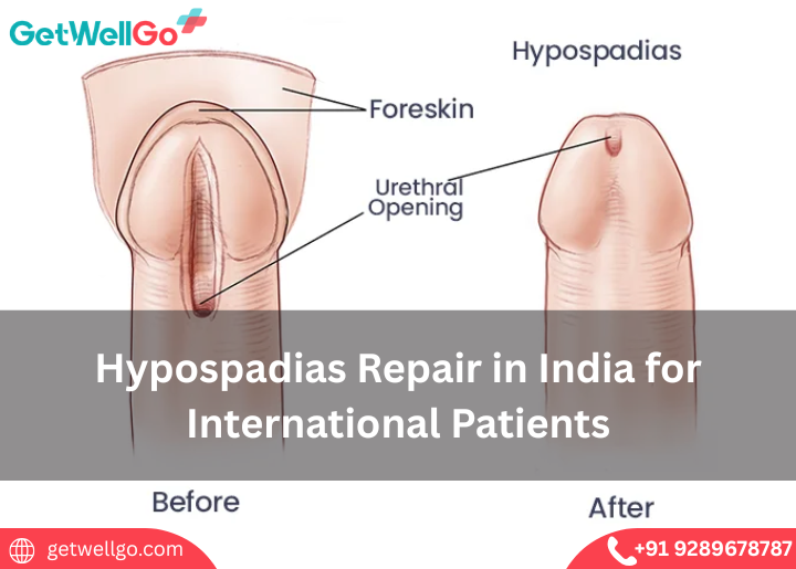
Hypospadias Repair in India ensures a normal future for your son. GetWellGo manages the expert pediatric surgery, family visas, and a safe, private hotel.
AV Fistula for Dialysis is your secure lifeline. We at GetWellGo handle your affordable vascular surgery abroad, travel logistics, and a safe healing stay.
Testicular Sperm Extraction in India helps build your family. GetWellGo discreetly arranges your top urologist, IVF clinic, and comfortable stay abroad.
Urethroplasty treatment in India restores comfort and urinary function. GetWellGo handles your expert urologist bookings, medical visas, and safe stay.
Retrograde Intrarenal Surgery in India removes kidney stones without cuts. GetWellGo handles your visa, hotel, and specialist care for a fast recovery.
Choose GetWellGo for Peyronie's disease treatment in India. Access skilled specialists, modern hospitals, cost-effective care, and full assistance for global patients.

.png)