
Historic Milestone: FMRI Celebrates 2500 Successful Bone Marrow Transplants
Celebrate a historic milestone of 2500 successful Bone Marrow Transplant at Fortis Gurgaon procedures, backed by our complete international patient visa care.
Read MoreHip dysplasia occurs when the hip joint bones don’t align properly. GetWellGo helps guide treatment for babies through bracing or surgery.

Hip Dysplasia by definition refers to a developmental abnormality of a joint and in particular hip dysplasia is characterized by an improperly formed hip joint leading to instability, pain and occasional arthritic changes. It commonly affects the joint where the ball (femoral head) nested in the socket (acetabulum) but is not smoothly and firmly inserted into the cup – like structure.
Hip dysplasia is of two broad categories:
There are several ways by which hip dysplasia can be triggered hence it may be genetic, environmental or both. Here it is some cases and factors, they are as follows:
Genetic Factors
Developmental Factors (DDH)
Trauma or Injury
Environmental Factors
Other Medical Conditions
Wear and Tear (Osteoarthritis): With age, the cartilage in Hip joint may steadily wear out thus developing or worsening of Hip Dysplasia results into pain and limited movements.
Obesity: Too much weight also creates on the hip joint brings possibility of development of dysplasia or increases the instability of the joint in case it already been affected.
Pre-existing Injuries: Any past hip injuries or hip replacement surgeries may have shifted the position/shape or form of the hip joint and may well lead to dysplasia.
The treatment of hip dysplasia depends with the degree of developing hip dysplasia, age, whether the hip dysplasia is congenital or not. Thus, some of the treatment methods include non-surgical ones and various surgical ones as well.
Non-Surgical Treatment
These are initial treatments that are usually given to patients who have trivial or first attack episodes or relatively young patients.
Surgical Treatment
Surgery might be advisable in extreme cases or where other treatments cannot help to treat the situation effectively.
Post-Surgical Rehabilitation
Rehabilitation is vital in the later stages to help the individual achieve functional recovery of the hip joint. This typically includes:
Long-Term Management
Adult hip dysplasia usually develops from hip dysplasia, which is more common in infancy or childhood, but left untreated or undiagnosed. In the long-run it would result in difficulties within the hip joint, problems like pain, instability, and even chances of developing osteoarthritis of the joint. Although hip dysplasia affects children more often, the disease may not manifest or progress in the adult population for many years.
The causes of hip dysplasia in adults are as follows:
Developmental dysplasia of hip or DDH refers to a condition in which the hip joint is not developed as it should in infants. The head of the thighbone is not firmly in place in the socket of the pelvis known as acetabulum. It can be partially or fully displaced or even totally displaced.
Newborn Physical Examination
Ultrasound Imaging
X-ray
Risk-Based Screening
Some of the patients might undergo imaging early even though the physical examination is normal due to certain risk factors that they might possess such as breech birth or family history.
Periacetabular Osteotomy (PAO)
Recovery:
Femoral Osteotomy
Hip Arthroscopy
This is suitable for: Weaker hip cases that have impingement, labral tear or cartilage problem.
What it does:
Open Reduction & Spica Casting (Pediatrics)
Total Hip Replacement (THR)
Considerations:
Hip dysplasia complications: In Infants and Children
In Teens and Adults
Celebrate a historic milestone of 2500 successful Bone Marrow Transplant at Fortis Gurgaon procedures, backed by our complete international patient visa care.
Read More
Experience advanced Corneal Collagen Cross-Linking in India to stabilize keratoconus and protect your sight, with full medical visa help from our travel team.
Read More
Experience the LINX Reflux Management System in India to stop severe heartburn using a tiny magnetic ring, fully organized by our fast-track medical visa team.
Read More
Experience advanced Stretta Procedure for GERD in India to eliminate chronic acid reflux without surgery, completely managed by our fast-track medical visa team
Read More
Get advanced vagus nerve stimulation treatment in India with expert care, modern hospitals, and affordable options for international patients.
Read More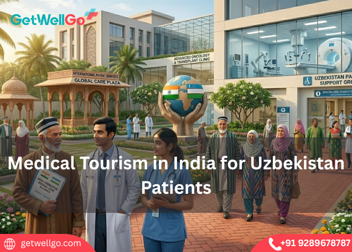
Medical tourism in India for Uzbekistan Patients made easy. GetWellGo handles your top-tier surgery, Uzbek translator, and a safe, warm home for your family.
Read More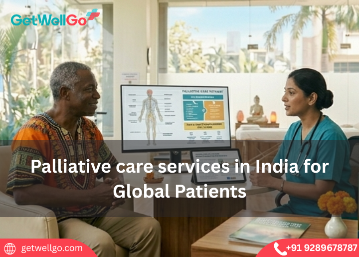
Palliative care services in India focus on your comfort and peace of mind. GetWellGo handles the expert pain relief, visa, and a serene, private home to heal.
Read More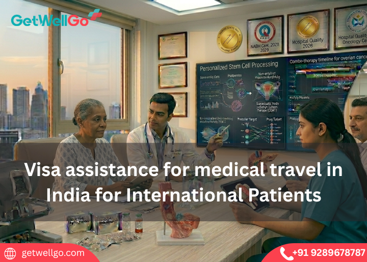
Get reliable visa assistance for medical travel to India. Support with documentation, approvals, and a smooth entry process for global patients.
Read More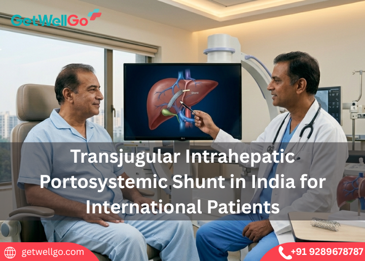
Transjugular Intrahepatic Portosystemic Shunt in India stops dangerous liver bleeding. GetWellGo handles your top specialist, visa, and a safe, cozy stay.
Read More
Keratoconus Treatment in India stops your vision from getting worse. GetWellGo handles your advanced eye care, visas, and a safe trip to save your sight.
Read More
Implantable Collamer Lens in India lets you ditch thick glasses forever. GetWellGo handles your expert eye doctor, flights, and a clear-vision holiday.
Read More
Cochlear Implants for Children in India opens a world of sound. We at GetWellGo plan your affordable ear surgery, airport pickup, and safe family trip.
Read More