An Atrial Septal Defect (ASD) is a congenital heart defect involving a hole in the wall (septum) between the heart's two upper chambers (atria). If the defect is large enough or is symptomatic, surgery or catheter-based treatment is typically needed to avoid complications such as heart failure, arrhythmias, or pulmonary hypertension.
Types of ASD Closure Procedures:
Catheter-Based Closure (Minimally Invasive)
Most common for secundum ASD
Procedure: A catheter is threaded through a vein in the groin and led to the heart. A closure device (such as an Amplatzer septal occluder) is inserted to close the hole.
Resume school/work: 1 week (assuming no complications)
Full recovery: 1–2 weeks
Recovery Tips:
Avoid heavy lifting for 1–2 weeks.
Bruising at the site of the catheter is normal.
Follow-up echo at 1 month to ensure device placement.
Blood-thinning drug (such as aspirin) may be used for 6 months.
Open-Heart ASD Surgery Recovery
Recovery Timeline:
ICU stay: 1–2 days
Total hospital stay: 5–7 days
Back to school/work: 4–6 weeks
Full physical recovery: 6–8 weeks
Sternum healing time: Up to 8–12 weeks (no contact sports until then)
Recovery Tips:
Avoid driving, strenuous activity, or lifting >5 kg for at least 6 weeks.
Incision care: keep dry and clean.
Breath-holding or physiotherapy exercises may be advised.
Follow-up visits to check heart rhythm and healing.
Minimally Invasive ASD Repair
Minimally invasive ASD repair is a new, low-risk method of repairing ASDs, traditionally performed using catheter-based device closure. It is best suited for secundum-type ASDs with good rim for secure anchoring of the closure device.
Key Features
Type: Catheter-based transcatheter device closure
Access site: Femoral vein (groin)
Incision: No chest incision; only a small groin puncture
Anaesthesia: Local with sedation or general (particularly in children)
Hospital stay: 1–2 days
Recovery time: 3–7 days
Procedure Steps
Imaging verification: TEE or intracardiac echo is performed before and during the procedure.
Catheter placement: A thin, flexible tube (catheter) is placed in the femoral vein and directed to the heart.
Device placement:
A self-expanding occluder device (e.g., Amplatzer Septal Occluder) is positioned across the defect.
It unfolds like two disks on either side of the septal hole and closes it.
Device release: After ascertaining proper position, the device is released from the catheter.
Closure: Catheter is withdrawn and a tiny dressing is applied over the puncture site.
ASD Patch Repair Procedure
ASD patch repair is a surgical technique applied to close larger or complicated atrial septal defects, particularly when catheter-based closure is not ideal. It's utilized most commonly in primum, sinus venosus, or large secundum ASDs.
When Is Patch Repair Indicated?
Large ASD (>38 mm)
Inadequate tissue rim for device deployment
Associated structural heart malformations (e.g., cleft in mitral valve)
Non-secundum ASDs (e.g., primum, sinus venosus)
Failed or contraindicated device closure
Step-by-Step: ASD Patch Repair Operation
Preoperative Preparation
Cardiac imaging: Echocardiography, TEE, ECG, blood work, occasionally cardiac MRI/CT
Stop food/water 6–8 hours prior to surgery
Consent and anaesthesiology assessment
Anaesthesia and Positioning
General anaesthesia
Patient positioned in a supine position (back lying)
Incision and Heart Exposure
A median sternotomy (down the center of the chest) incision is created
The breastbone is split to expose the heart
Heart is attached to a cardiopulmonary bypass (CPB) machine, which replaces the heart and lung function during surgery
Accessing the Defect
The right atrium is opened to see the atrial septal defect directly
Patch Closure of the Defect
The hole is closed by a patch. Patch materials used are:
Dacron (man-made material)
Autologous pericardium (patient's own heart covering)
Bovine pericardium (biological)
The patch is sewn over the defect with fine sutures
Closure and Weaning Off Bypass
Heart is closed and up and running
CPB is being stopped
Temporary pacing wires or chest drains can be inserted
Chest Closure
The sternum is wired back together
Soft tissues and skin are sutured
ASD heart defect surgery risks
Following is a comprehensive summary of risks and possible complications of Atrial Septal Defect (ASD) heart defect surgery, for open-heart surgical repair (patch closure) and catheter-based (device closure).
ASD surgery complications (Both Surgical and Device Closure):
Arrhythmias
Residual ASD leak
Infection
Stroke
Device-related complications
Pericardial effusion
Blood clots
Atrial Septal Defect Surgical Outcomes
Atrial Septal Defect (ASD) closure via open-heart surgery (patch repair) or catheter-based device closure has great results if done in timely, proper cases. Here's a complete analysis of the anticipated results, long-term advantages, and success factors.
Success Rate
Overall success rate: 95%–98%
Complete closure without any residual shunt in most patients
Normal life expectancy can be anticipated after closure in the majority of cases
Symptom Relief
Fatigue, breathlessness: Relief within weeks
Frequent respiratory infections (in children): A significant reduction
Palpitations: Reduces, although arrhythmias can still exist in some adults
Exercise intolerance: Severe improvement within 2–3 months
Enlargement of the heart (RA/RV): Gradually reverses after closure
Long-Term Results
Cardiac function: Normalizes within 6–12 months in most patients
Pulmonary pressure: Pulmonary hypertension can improve or remain stable if treated early
Risk of stroke: Significantly decreased after closure
Pregnancy: More secure after closure; many women are able to have full-term pregnancies
Life expectancy: Almost-normal if ASD closed before complications arise
Pediatric Results
Children usually recover more quickly and adapt better long-term
Growth and development adapt post-surgery
Most are able to engage in full physical activity after 6–8 weeks
Adult & Elderly Outcomes
Results are still great but:
Certain arrhythmias (such as atrial fibrillation) can remain
If ASD is closed beyond age 40–50, recovery of heart function/size can take longer
Adults with pulmonary hypertension that has not been treated may not gain full benefit
ASD operation recovery tips
Here are recovery tips that are vital following Atrial Septal Defect (ASD) surgery, tailored to both open-heart (patch repair) and catheter-based (device closure) surgery:
ASD Surgery Recovery Tips
Following Open-Heart ASD Patch Repair:
Rest: Rest is imperative for 4–6 weeks. Refrain from stairs and heavy exercise initially.
Wound care: Keep the chest wound clean and dry. Monitor for infection (redness, pus, fever).
Pain management: Take prescribed drugs. Chest pain, particularly on coughing or movement, is expected.
Sleep position: Sleep slightly above flat (2–3 pillows). Avoid flat sleeping for the first few days.
No lifting: Do not handle objects >5 kg for 6–8 weeks (to avoid straining the healing sternum).
No driving: At least 4–6 weeks after surgery and only after clearance by your doctor.
Pulmonary exercises: Employ spirometer (if provided) to clear lungs and avoid pneumonia.
Diet: Low-sodium, light diet. Fiber-rich foods prevent constipation related to pain medication.
Watch for symptoms: Report shortness of breath, chest pain, dizziness, or wound discharge.
Following Catheter-Based ASD Closure (Minimally Invasive):
Restriction of activity: Light activity for 1–2 days. Heavy lifting or strenuous exercise should be avoided for 1–2 weeks.
Groin care: Bandage kept dry for 24 hours. Bruising or soreness at catheter site is mild.
Hydration: Drink lots of water to clear contrast dye administered during the procedure.
Medication: Aspirin or antiplatelet drug is generally given for 6 months. It should be taken daily.
Echo follow-up: Performed at 1–3 months to verify device placement and closure
Prevent dental treatment: Postpone any dental treatment by 6 months to decrease the chance of infection around the device.
Atrial Septal Defect Post-op Care
Successful post-op care is crucial for safe healing and avoiding complications, and restoring normal activity—regardless of whether the ASD was closed by open-heart surgery or catheter-based device closure.
Wound & Incision Care
Open-Heart Surgery:
Maintain incision clean and dry (chest and possibly leg if vein used).
Do not apply ointments or powders unless ordered by physician.
No soaking in bathtub or swimming pool for at least 4 weeks.
Inspection daily for redness, discharge, swelling, or wound separation.
Catheter-Based Closure:
Groin puncture care:
Wear dressing for 24–48 hours
Monitor for swelling or bleeding
Avoid excessive bending or squatting for a few days
Heart & Circulatory Care
Medications:
Blood thinners such as aspirin are commonly prescribed for 3–6 months (device closures).
Anti-arrhythmics might be employed in certain adult patients.
Monitor pulse rate and rhythm: Report palpitations or an increased heartbeat.
Follow-up echocardiograms are crucial to determine:
Device position (if used)
Integrity of closure
Recovery of heart chamber
Activity & Mobility
Light walking
Open-heart: After 3–5 days
Device Closure: After 1–2 days
Stairs
Open-heart: After 2 weeks
Device Closure: After 3–4 days
Driving
Open-heart: After 6 weeks
Device Closure: After 1–2 weeks
Heavy lifting
Open-heart: After 8–12 weeks
Device Closure: Avoid for 2 weeks
Sports/exercise
Open-heart: After 3 months (doctor clearance)
Device Closure: After 1 month (light exercise)
Sleep & Rest
Sleep with 2–3 pillows for elevation (first 1–2 weeks)
Avoid sleeping on your stomach
Fatigue is usual for 4–6 weeks—balance rest with gradual activity
Nutrition & Hydration
Eat small, balanced meals: Fruits, vegetables, lean protein
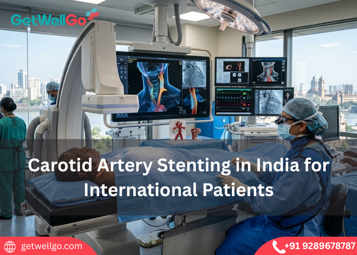
Restore safe brain blood flow with Carotid Artery Stenting in India organized smoothly by our team, from rapid specialist booking to a cozy recovery stay.
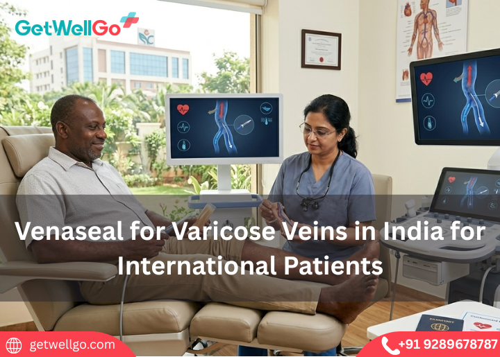
Experience advanced Venaseal for Varicose Veins in India to seal damaged veins with medical glue, fully managed by our fast-track medical visa travel team.
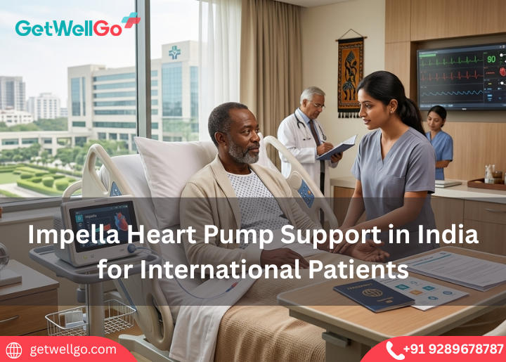
Protect failing heart function with Impella Heart Pump Support in India organized smoothly by our team, from emergency hospital booking to a safe recovery.
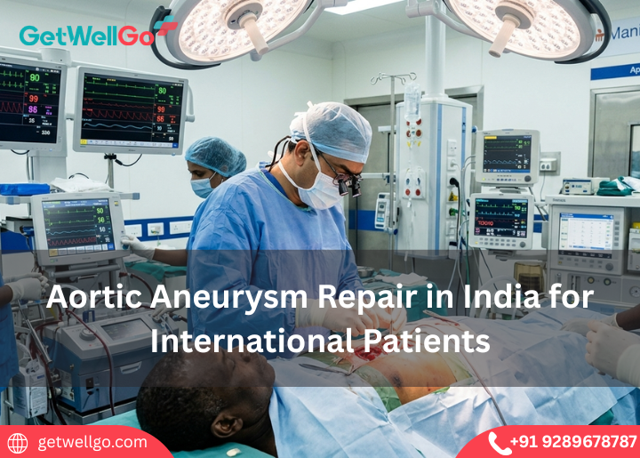
Aortic Aneurysm Repair in India: Fix weak artery walls safely before they rupture. We connect you to elite vascular teams, handle fast visas & a quiet stay.
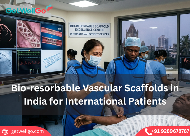
Bio-resorbable Vascular Scaffolds in India: Open your arteries with a stent that melts away naturally! We arrange top cardiologists, visas & a cozy stay.
Shockwave Lithotripsy for Arteries in India: Clear hard calcium gently with sonic waves! We arrange your top cardiologist, medical visa & a cozy recovery stay.
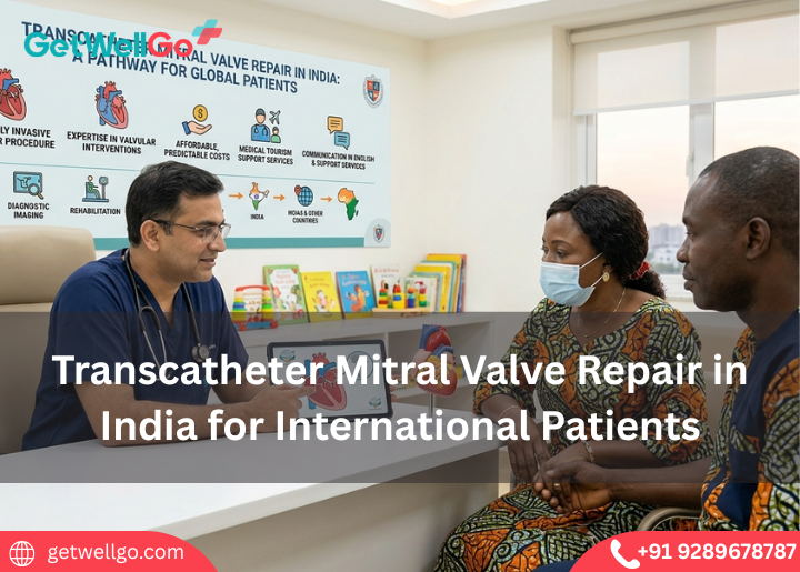
Explore safe, minimally invasive mitral valve repair in India for global patients with expert care, modern hospitals, and cost-effective treatment options.
Choose GetWellGo for Transcatheter Aortic Valve Replacement in India. International patients receive expert cardiac care, modern hospitals, and cost savings.
Choose GetWellGo for pediatric cardiac catheterization in India. International patients get advanced care, skilled pediatric cardiologists, and cost-effective treatment.
MitraClip Procedure in India fixes leaky heart valves without open surgery. GetWellGo handles your top cardiac clinic, medical visa, and safe recovery.
Endovascular Aneurysm Repair in India saves lives without major open surgery. GetWellGo handles your top vascular clinic bookings, visas, and safe recovery.
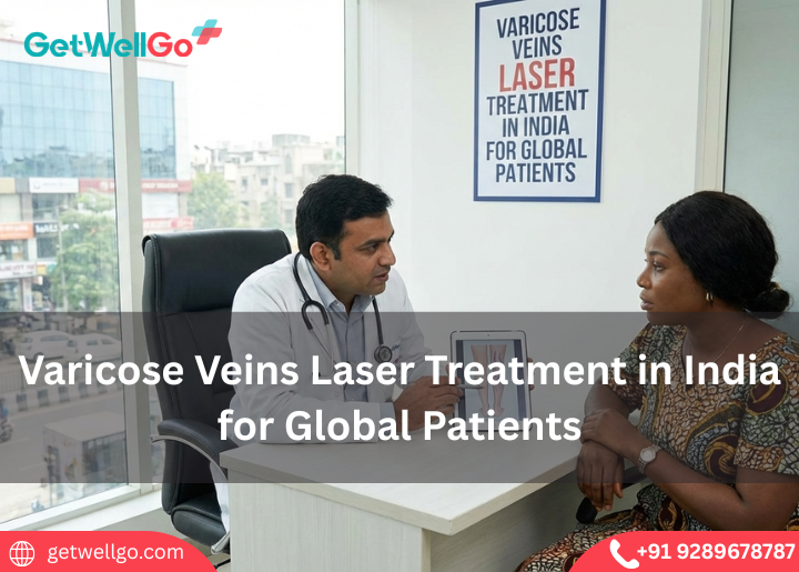
Get world-class varicose veins laser treatment in India at competitive costs, combining advanced technology, quick recovery options, and smooth travel support for global patients.

.png)