Historic Milestone: FMRI Celebrates 2500 Successful Bone Marrow Transplants
Celebrate a historic milestone of 2500 successful Bone Marrow Transplant at Fortis Gurgaon procedures, backed by our complete international patient visa care.
Read MoreLearn how a radiologist plays a vital role in diagnosing and guiding treatment for laryngeal cancer with expert support from GetWellGo’s medical team.

Laryngeal cancer is a type of cancer that affects the larynx or the human voice box that exists in the throat. It is involved in phonation, as well as in respiration or swallowing processes. Laryngeal cancer is the cancer that starts from the squamous cell carcinoma that is located in the lining of the larynx.
Radiology is another important aspect when it comes to laryngeal cancer diagnosis and staging, as well as planning of the right treatment. Major imaging examinations that are used in the diagnosis of laryngeal cancer are CT scans, MRI, and PET scan.
MRI is especially beneficial for the evaluation of soft tissues; it may give better visions of the larynx and the adjacent structures than CT. It is also used in the assessment of the following:
MRI provides better contrast of soft tissues than the CT and it can determine the location of the tumor in close proximity to crucial structures.
CT scans give good images of the larynx, throat, and other related areas and aid in determining degree of tumor infiltration of the affected tissues. The CT scan can reveal:
Contrast-enhanced CT scans are then more useful to define the tumor and especially the anatomy of the surroundings.
The choice of the imaging technique in laryngeal cancer is dependent with the question asked and the stage or area of the cancer. However, several imaging methods may be employed to provide detailed information on the type of tumor, its size and the relation with the surrounding tissues. The following are the useful imaging modalities that are useful in the management of laryngeal cancer and the benefits of each:
Key Aspects of CT Imaging in the Diagnosis of Laryngeal Cancer:
Under the radiology of laryngeal tumors includes methods that aid in the diagnosis, determining the type of tumors, planning a treatment process as well as monitoring the progress of the tumor within the larynx.
Radiology is important because the larynx is a three-dimensional structure and the tumor’s scope cannot be evaluated during endoscopy.
Check localized tumors before they become malignant; extend their probes to other tissues or part of the body.
This increases an opportunity of voice preservation, decreased invasive procedures and better survival rates.
MRI
CT
High-Resolution Endoscopy (Video Laryngoscopy)
PET-CT
|
Feature |
MRI |
CT |
|
Soft Tissue Contrast |
Excellent (best for early tumor detection, soft tissue detail) |
Good, but less than MRI |
|
Cartilage Invasion Detection |
Very good (especially early cartilage infiltration) |
Very good for advanced cartilage destruction (erosion, sclerosis) |
|
Bone Involvement |
Less sensitive than CT for bony structures |
Best for assessing bone erosion and thyroid cartilage ossification |
|
Lymph Node Evaluation |
Good (can show node size and necrosis) |
Very good (better for detecting calcified nodes) |
|
Speed |
Slower (30–45 min scan) |
Faster (5–10 min scan) |
|
Availability & Cost |
More expensive, less available |
Widely available, less costly |
|
Artifacts |
Motion sensitive (swallowing can blur images) |
Less sensitive to motion artifacts |
|
Use in Post-treatment Follow-up |
Superior for differentiating recurrence vs post-radiation fibrosis |
Less specific for fibrosis vs recurrence |
|
Need for Contrast |
Gadolinium contrast often needed |
Iodinated contrast usually needed |
|
Contraindications |
Pacemaker, severe claustrophobia, metal implants |
Generally safe for most patients |
Mass or Tumor
Asymmetry
Mucosal Thickening
Loss of Normal Fat Planes
Vocal Cord Fixation
Cartilage Changes
Sclerosis (early invasion)
Airway Narrowing
Lymphadenopathy
Enhancement Patterns
Subglottic Extension
Involvement of Adjacent Structures
Laryngeal cancer staging depends on:
Radiologic T-Staging of Laryngeal Cancer
Radiologic N-Staging (Nodes)
Radiologic M-Staging (Distant Metastasis)
Ultrasonography can be used as a modality for assessment of neck lymph nodes. It can also differentiate between benign and malignant nodes, aid in decision-making regarding biopsy in an axillary node.
Main Uses of Ultrasound in Laryngeal Cancer:
Celebrate a historic milestone of 2500 successful Bone Marrow Transplant at Fortis Gurgaon procedures, backed by our complete international patient visa care.
Read More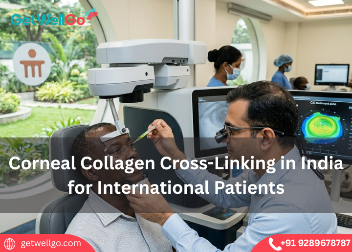
Experience advanced Corneal Collagen Cross-Linking in India to stabilize keratoconus and protect your sight, with full medical visa help from our travel team.
Read More
Experience the LINX Reflux Management System in India to stop severe heartburn using a tiny magnetic ring, fully organized by our fast-track medical visa team.
Read More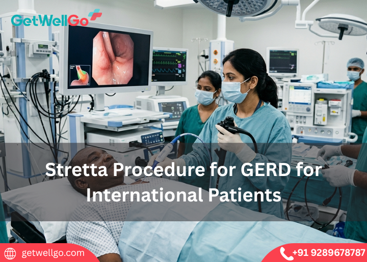
Experience advanced Stretta Procedure for GERD in India to eliminate chronic acid reflux without surgery, completely managed by our fast-track medical visa team
Read More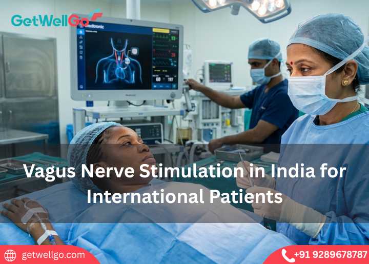
Get advanced vagus nerve stimulation treatment in India with expert care, modern hospitals, and affordable options for international patients.
Read More
Medical tourism in India for Uzbekistan Patients made easy. GetWellGo handles your top-tier surgery, Uzbek translator, and a safe, warm home for your family.
Read More
Palliative care services in India focus on your comfort and peace of mind. GetWellGo handles the expert pain relief, visa, and a serene, private home to heal.
Read More
Get reliable visa assistance for medical travel to India. Support with documentation, approvals, and a smooth entry process for global patients.
Read More
Transjugular Intrahepatic Portosystemic Shunt in India stops dangerous liver bleeding. GetWellGo handles your top specialist, visa, and a safe, cozy stay.
Read More
Keratoconus Treatment in India stops your vision from getting worse. GetWellGo handles your advanced eye care, visas, and a safe trip to save your sight.
Read More
Implantable Collamer Lens in India lets you ditch thick glasses forever. GetWellGo handles your expert eye doctor, flights, and a clear-vision holiday.
Read More
Cochlear Implants for Children in India opens a world of sound. We at GetWellGo plan your affordable ear surgery, airport pickup, and safe family trip.
Read More