Urology
Ureterocalycostomy
Ureterocalycostomy
Get expert ureterocalycostomy surgery with GetWellGo. Trusted hospitals, top urologists & complete care for international patients. Book your treatment today.
Ureterocalycostomy Surgery
Ureterocalycostomy is a reconstructive urological surgery done to help in the restoration of the blood flow of urine between the ureter, the tube that transfers the urine of the kidney into the bladder, and the renal calyx, a portion of the collecting system of the kidney. It is commonly performed in circumstances where the routine like pyeloplasty cannot be performed or has failed.
What is Ureterocalycostomy?
Ureterocalycostomy is a surgical procedure where the ureter is joined to a lower pole renal calyx when the supply is blocked because of the removal of the obstructed segment of the renal pelvis. It establishes a pathway of a urinary outlet of the kidney into the ureter.
This surgery is believed to be a salvage or re-do operation in complex ureteropelvic junction obstruction (UPJO) or following failure of pyeloplasty.
Advantages of Ureterocalycostomy.
- Offers an alternative drainage route in case of failure by the conventional alternatives.
- Enhances renal functions in salvage kidneys.
- Minimally invasive (laparoscopic or robotic) can be done.
Ureterocalycostomy surgery procedure
Ureterocalycostomy is a connective urological surgery involving direct communication between the ureter and the lower calyx of the renal to bypass a non-dilated or scarred ureteropelvic junction (UPJ). Its primary implementation, as a salvage procedure, occurs after the failure of a pyeloplasty, or in cases where the renal pelvis is intrarenal and inoperable.
Ureterocalycostomy Step by Step Procedure:
Preparation before Surgery
Investigations:
- Determination of the functioning of renal activity and the obstruction: Ultrasound, IVP (Intravenous Pyelogram), CT Urogram or MAG3/ DTPA renal scan.
- Antibiotic Treatment: This is a preoperative measure in the prevention of infection.
- Anaesthesia: Performed by general anaesthesia.
Positioning:
-
The patient is put either in flank or lateral decubitus (based on the type of surgery).
Surgical Approaches
Ureterocalycostomy may be carried out with the following:
- Open surgery (conventional method)
- Laparoscopic surgery
- Robotic-aided surgery (more accurate and better sight)
The process involved in all the three procedures is largely the same with the only few differences in the incision size and equipment.
Kidney and Ureter Exposed
- Open surgery involves a flank/subcostal incision, whereas laparoscopy/robotics involve small port incisions.
- Careful exposure of the kidney and the proximal ureter is done by dissecting through the perinephric fat.
- The ureteropelvic junction (UPJ) and renal pelvis are located.
Resection of the Obstructed Segment
- The hampered UPJ as well as scar tissue is removed in totality.
- The ureter is excised until healthy and viable tissue is observed to have good vascularity.
The choice and preparation of Calyx
- The kidney forms a lower pole which is known because it offers the most dependent point of drainage.
- To open the lower calyx, a small piece of renal parenchyma (1-2 cm) is removed.
- Control of bleeding is done with the use of bipolar cautery or fine sutures.
Mobilisation of Ureter
- The ureter is manipulated enough to get to the lower calyx without strain.
- In case of need, the ureter is spatulated (cut longitudinally) at its terminal to increase the anastomotic surface.
Uretero-Calyceal Anastomosis
- Anastomosis of the ureter to the calyceal opening is done with a fine absorbable suture (e.g., 5-0 Vicryl or 6-0 Vicryl).
- The posterior wall is first sutured and then the anterior wall and this gives a watertight connection.
Stent Placement
- To ensure patency and wound healing, a DJ (Double J) ureteral stent is placed across the anastomosis.
- The stent runs out of the renal calyx into the bladder.
Drainage and Closure
- A perinephric drain is inserted in the area of the anastomosis to identify leakage of the urine.
- The wound is sutured (muscle, fascia, skin).
Postoperative Care
- Length of Hosptialization: 3-7 days.
- Drain Removal: Typically this happens after 48-72 hours when the drainage has subsided.
- Stent Removal: 4-6 weeks of cystoscopy.
- Follow-up Imaging: Ultrasound or renal scan (MAG3) is followed after the removal of the stents to ensure that the urine is flowing freely.
Success Rate
- Open surgery: 85–90% success
- Laparoscopic/robotic: 85 95% success.
- The definition of success is based on the better drainage, relief of symptoms, and renal stability.
Indications for ureterocalycostomy
Ureterocalycostomy is a reconstructive urological surgery mainly used to treat complex or recurrent ureteropelvic junction obstruction (UPJO) in those cases in which the standard reconstructive procedures like pyeloplasty are not possible or unsuccessful. It is a salvage operation to maintain kidney functioning in case of failure of other operations.
The primary signs are the following:
Unsuccessful or Recurrent UPJ Obstruction
- Most common indication.
- In the event of primary pyeloplasty or endopyelotomy failure because of fibrosis, scarring, or recurrent obstruction of the UPJ.
- The ureterocalycostomy creates a new pathway of drainage by draining the ureter directly to a lower pole calyx.
Small Renal Pelvis or intrarenal
- In cases of a small or deeply rooted renal pelvis (intrarenal pelvis) which makes it technically difficult to carry out normal pyeloplasty.
- A calyx anastomosis provides a superior and more reliable drain out.
High Ureteral Insertion
- In cases where the ureter is inserted high within the renal pelvis resulting in poor dependent angle of drainage.
- This is repaired by ureterocalycostomy which adjoins the ureter to a lower more gravity dependent calyx.
Periureteral or widespread Peripelvic Fibrosis
- Observed in reoperative cases or chronic inflammation, or fibrosis of the retroperitoneal tissue, tissue planes are scarred and dissection along the renal pelvis is dangerous.
- The lower calyx provides a healthy and safe position of anastomosis.
UPJ Iatrogenic or Traumatic Injury
- When there is surgical trauma to the UPJ in the course of past operations (e.g., stone surgery, vascular surgery, or renal trauma).
- In case of an extensive damage of the proximal ureter or renal pelvis, ureterocalycostomy can be used to re-establish drainage.
Long Upper Ureteric Strictures
- In cases where the stricture is located in the upper ureter and proceeds near the renal pelvis leaving a shortage of length to carry out a standard pyeloplasty.
- The process circumvents the pathogenic area.
Renal Abnormal or Deformed Anatomy
Conditions such as:
- Horseshoe kidney
- Malrotated kidney
- Ectopic kidney
- Post-inflammatory changes
These can abnormally shape the normal renal pelvic structure, thus complicating conventional reconstruction.
Pediatric Indications
-
Ureterocalycostomy may be viewed as a salvage procedure that may preserve renal function in children who have failed primary pyeloplasty or poor renal pelvis development.
Other Rare Indications
- Post tubercular scarring of UPJ.
- After open or laparoscopic surgery: determine the history of injury.
- A very rare indication: Renal pelvis agenesis or atresia.
Ureterocalycostomy recovery time
The success of recovery following ureterocalycostomy is dependent on the type of surgery (open, laparoscopic, or robotic), general health of the patient and kidney functionality prior to surgery.
Generally, healing is slow and positive, particularly by using the least invasive techniques.
Postoperative Period (Hospital Stay)
-
Duration: 3 to 7 days
Monitoring:
- Close monitoring is paid to urine output and drain output.
- Opioid medication and IV antibiotics are administered.
- A urinary catheter is retained between 1 and 2 days.
- A perinephric drain (near the surgical site) is discharged as soon as the drainage reduces.
- Mobilization: A majority of patients begin to walk 1-2 days (sooner laparoscopic or robotic surgery) after surgery.
Early Recovery (First 2–4 Weeks)
At Home:
- Post-discharge light activities are permitted.
- Do not over exert one.
- Eat well and stay hydrated to facilitate urine flow and avoid infection.
Symptoms:
- Light discomfort in the flank or back pain lasting 1-2 weeks is normal.
- The DJ stent may cause some urgency or burning sensation in the urine.
Follow-up:
- Initial examination is typically after 7 -10 days.
- Stitches (where used) taken out at 10-14 days.
Stent Removal Period
- Timing: 4-6 weeks (removal after surgery).
- DJ stent that is inserted during the surgery is taken out through a cystoscopy procedure.
- The occurrence of mild burning or frequency of urination can happen 1-2 days after the procedure by the time the stents are removed.
Complete Recovery
- Open surgery: 6–8 weeks
- Laparoscopic surgery: 3–4 weeks
- Robotic surgery: 2–3 weeks
By this time:
- Wounds are fully healed.
- Normal levels of activity are restored.
- Urinary and pain symptoms are completely resolved.
Follow-Up
Imaging such as Renal Scan or Ultrasound:
- Conducted at 6 -8 weeks and 3-6 months after surgery.
- Resolves the obstruction of urine and ensures that urine flows freely.
- Kidney functional tests: Routine observation of renal stability.
Open ureterocalycostomy surgery
- Open ureterocalycostomy is a type of reconstructive urologic surgery that is done to reconstruct urinary drainage of the kidneys to the ureter by providing a direct interrelation between the ureter and the lower renal calyx.
- It is commonly pointed out when conventional interventions such as pyeloplasty do not or cannot be effective or the renal pelvis is small, intrarenal or scarred.
- This is an open procedure whereby the surgeon has a direct view of the surgery and this is helpful in complex or reoperative cases.
Objectives of the Surgery
- To avoid an obstructed ureteropelvic junction (UPJ).
- To differentiate a new, dependent drainage pathway of urine.
- To maintain the function of a kidney that would otherwise not be able to persist with its functioning as a result of continuous obstruction.
Indications
Open ureterocalycostomy is done on cases like:
- Recurrent UPJ obstruction (Failed pyeloplasty).
- Intrarenal small or pelvis renal.
- Unable to do standard pyeloplasty because of high ureteral insertion.
- Peripelvic fibrosis or scarring of UPJ.
- Long upper ureteric stricture for close to the renal pelvis.
- Iatrogenic or traumatic UPJ injury.
Factors Affecting Ureterocalycostomy cost India
The following are the key factors that will determine the amount you will pay (or at least budget) in having ureterocalycostomy in India:
Complexity of the Case
- History of past surgeries, scarring/fibrosis, anatomic deviation (e.g. intrarenal pelvis) increases length of surgery, risk, resource usage.
- More complex = higher cost.
- Hypothesis: In other urology procedures, complexity makes cost high.
- On iterative or salvage surgeries (most ureterocalycostomies are) charge more.
Kind of Surgical Methodology and Technology
- Minimally invasive surgery (laparoscopic or robot) or open (standard) cost. Minimally invasive usually is more expensive due to specialised equipments, long operating period, highly sophisticated instruments.
- Even though in one instance the government scheme charges the same price between open and laparoscopic, in the private hospitals it is better to have a premium price billed to laparoscopic/robotic.
- Consumable costs with high prices, stents, advanced imaging, etc are additional costs.
Hospital / Facility Type & Infrastructure
- Premium private hospitals (more so in metros) are expensive as compared to smaller hospitals or those located in smaller cities.
- Accreditation, intensive care unit availability, specialised unit, foreign patient services are additional expenses.
- Location issues: high overheads (property, staffing, utilities) in a metropolitan city.
Experience of Surgeon / Specialist Group
- The prices of highly skilled urologists who perform complicated reconstructive surgeries might be higher.
- Depending on the case, a team (urologist + nephrologist + anaesthetist + possibly vascular surgeon) can add to the cost of the complex cases.
Length of Stay /Postoperative-care
- Cost is increased by longer ICU stay, complications, further procedures (e.g., drainage, redo procedures) etc.
- Pre-operative tests + imaging and follow-up are also external contributors.
Geographical Location
- The price differs according to the urban areas
- Also the cost differs between private and public hospitals.
International Patient Add-ons
- In the case of foreign patients or expatriates: the extra expenses can be paid to translators, airport/hospital transportation, non-hospital accommodation, visa/immigration process, and additional diagnostics.
- It should not be included in the surgery package and should be accounted independently.
Pre-existing Condition and Risk Profile
- In case of comorbidities (e.g., renal impairment, obesity, cardiovascular disease) in a patient, there is an increase in risk, which may require more resources, monitoring, which is more expensive.
- Owing to increases of cost in re-operative fields (past surgery).
Food and Devices to be implanted
- Other cost elements include the use of stents, drainage catheters, special sutures, specialised devices (where necessary) and so on.
- The price of ureterocalycostomy might not depend significantly on the price of the implants, but consumables do.
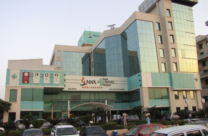
Best hospital for ureterocalycostomy India
- Artemis Hospital, Gurgaon
- Medanta-The Medicity, Gurgaon
- Fortis Memorial Research Institute, Gurgaon
- Max Hospital, Saket
Conclusion
Ureterocalycostomy is a serious reconstructive urological operation aimed at re-establishing the urinary drainage through directly connecting the ureter to the lower renal calyx in place of an obstructed ureteropelvic junction (UPJ). It is a salvage operation in complicated or recurrent cases when other methods, including pyeloplasty cannot be performed on or have failed. The open ureterocalycostomy method is a highly effective technique with high success rates in the long term (85-90%), and it does not affect the functioning of the kidneys. Recovery can be expected to last 68 weeks, and full healing and recovery of urinary flow can be achieved after the removal of the stents. Although the process is rather technical, results are highly positive provided that it is conducted by competent urologic surgeons.
Ureterocalycostomy surgery India GetWellGo
GetWellGo is regarded as a leading supplier of healthcare services. We help our foreign clients choose the best treatment locations that suit their needs both financially and medically.
We offer:
- Complete transparency
- Fair costs.
- 24 hour availability.
- Medical E-visas
- Online consultation from recognized Indian experts.
- Assistance in selecting India's top hospitals for Ureterocalycostomy treatment.
- Expert urosurgeon with a strong track record of success
- Assistance during and after the course of treatment.
- Language Support
- Travel and Accommodation Services
- Case manager assigned to every patient to provide seamless support in and out of the hospital like appointment booking
- Local SIM Cards
- Currency Exchange
- Arranging Patient’s local food
FAQ
1. Is ureterocalycostomy a possibility with international patients?
- Yes. Most of the accredited Indian hospitals have international patient departments which offer visa assistance, airport transfers, translators and follow-ups. India is also reputable in good quality reconstructive urology treatment at half the price of the west.
2. What is the prognosis of the surgery in the long-term?
- The majority of patients have stable urine drainage and functioning of kidneys following the surgery. A follow-up analysis like ultrasound or renal scans should be done frequently to detect any recurrence in time.
3. Is it possible to repeat the surgery in case of failure?
- Whereas ureterocalycostomy is typically a definitive (final) treatment, on exceptional incidences of renal recurrence additional corrective measures like redo anastomosis or renal autotransplantation can be taken into consideration.
TREATMENT-RELATED QUESTIONS
GetWellGo will provide you end-to-end guidance and assistance and that will include finding relevant and the best doctors for you in India.
A relationship manager from GetWellGo will be assigned to you who will prepare your case, share with multiple doctors and hospitals and get back to you with a treatment plan, cost of treatment and other useful information. The relationship manager will take care of all details related to your visit and successful return & recovery.
Yes, if you wish GetWellGo can assist you in getting your appointments fixed with multiple doctors and hospitals, which will assist you in getting the second opinion and will help you in cost comparison as well.
Yes, our professional medical team will help you in getting the estimated cost for the treatment. The cost as you may be aware depends on the medical condition, the choice of treatment, the type of room opted for etc. All your medical history and essential treatment details would be analyzed by the team of experts in the hospitals. They will also provide you with the various types of rooms/accommodation packages available and you have to make the selection. Charges are likely to vary by the type of room you take.
You have to check with your health insurance provider for the details.
The price that you get from GetWellGo is directly from the hospital, it is also discounted and lowest possible in most cases. We help you in getting the best price possible.
No, we don't charge patients for any service or convenience fee. All healthcare services GetWellGo provide are free of cost.